The dental microscope helps identify even the smallest canals or cracks, leading to a more thorough and successful treatment.
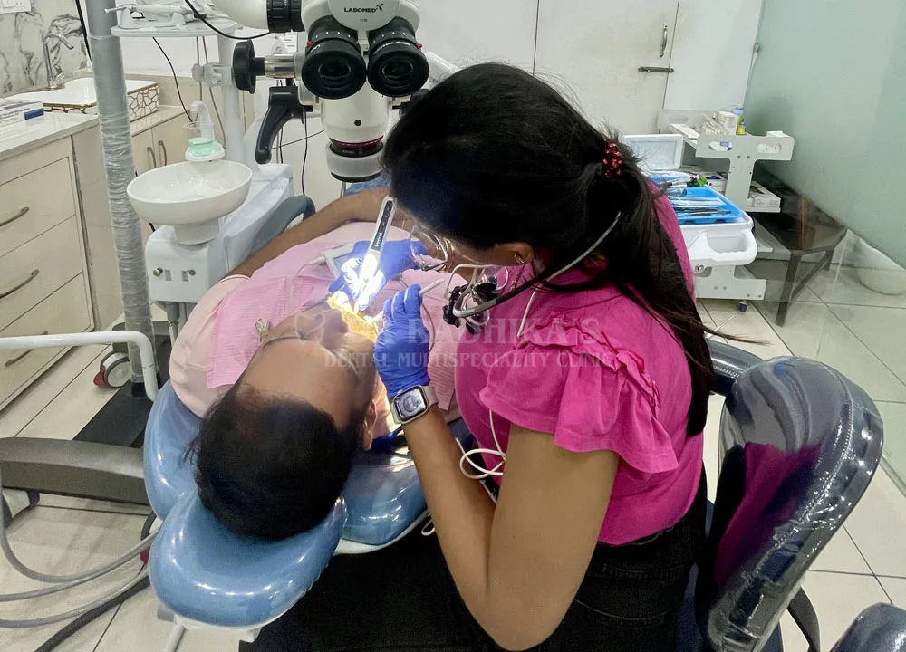
At Dr. Radhika Multispecialty Clinic, we are proud to offer Microscopic Root Canal Treatment—a modern, advanced, and highly precise approach to saving damaged or infected teeth. Unlike traditional root canal therapy, microscopic root canal treatment uses a dental microscope to enhance visibility and accuracy, ensuring better outcomes, minimal discomfort, and long-term dental health.
What Is Microscopic Root Canal Treatment?
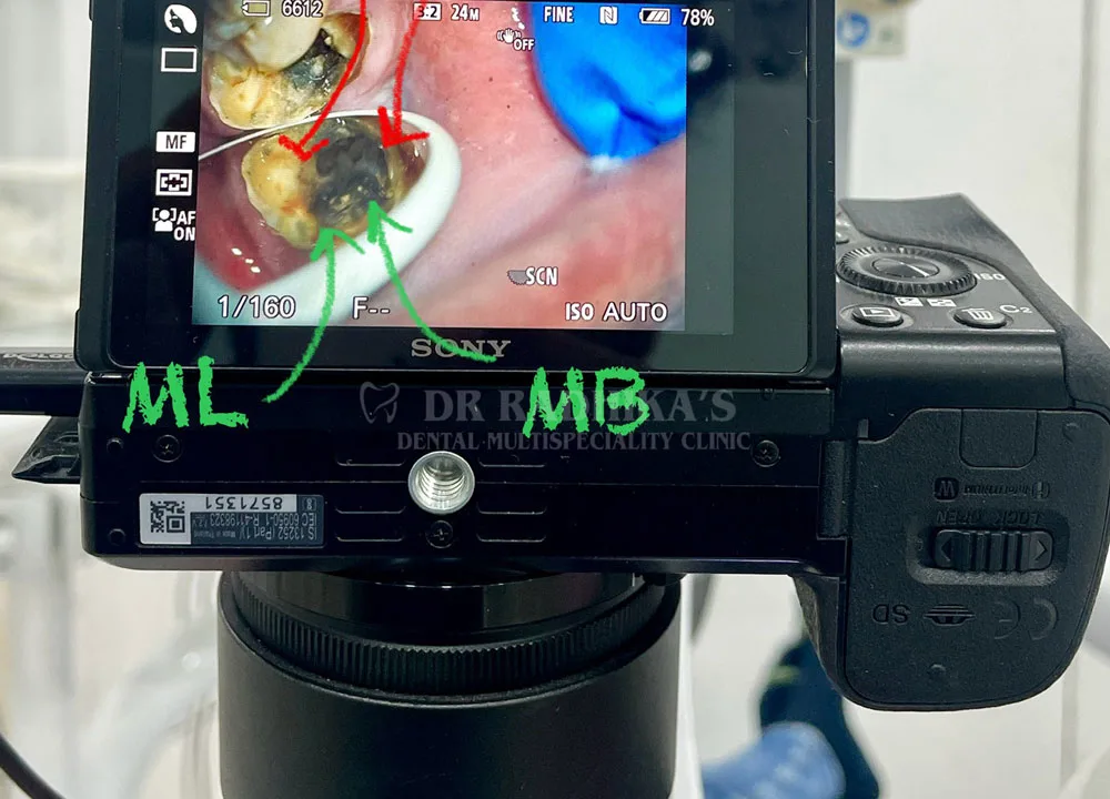
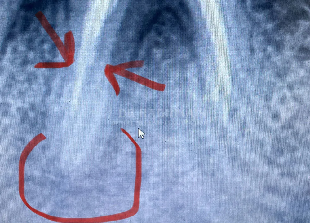
Microscopic root canal treatment is an advanced endodontic procedure that involves the use of a high-powered dental microscope to visualize the interior of the tooth. The magnification (typically 3x to 20x) and focused illumination provided by the microscope allow the dentist to detect tiny details such as hidden canals, fine cracks, or accessory canals that are often missed with the naked eye.
This technology significantly improves the chances of successful root canal therapy by ensuring complete cleaning, shaping, and sealing of the root canals.
Why You Might Need a Root Canal
A root canal is typically required when the pulp inside the tooth becomes infected or inflamed due to deep decay, trauma, cracks, or repeated dental procedures. If left untreated, this can lead to severe pain, swelling, and eventually tooth loss. Symptoms that may indicate the need for a root canal include:
Persistent toothache or pain when chewing
Sensitivity to hot or cold
Swelling or tenderness in nearby gums
Discoloration or darkening of the tooth
Presence of pus or abscess
Advantages of Microscopic Root Canal Treatment
Greater Precision The dental microscope helps identify even the smallest canals or cracks, leading to a more thorough and successful treatment.
Improved Success Rate With enhanced visibility, the chances of leaving behind infected tissue are greatly reduced, lowering the risk of future infections.
Conservative Approach The precision of the microscope allows dentists to remove less tooth structure, preserving more of your natural tooth.
Comfort and Safety Advanced tools and techniques ensure a pain-free experience, often completed in a single visit.
Better Diagnosis Cracks and complex canal structures are more easily detected, which helps in planning the most effective treatment strategy.
Our Expertise in Microscopic Endodontics
At Dr. Radhika Multispecialty Clinic, our team of experienced endodontists is trained in the use of state-of-the-art dental microscopes and rotary instrumentation. We follow strict sterilization protocols and use biocompatible materials to ensure that your tooth remains healthy and functional for years to come.
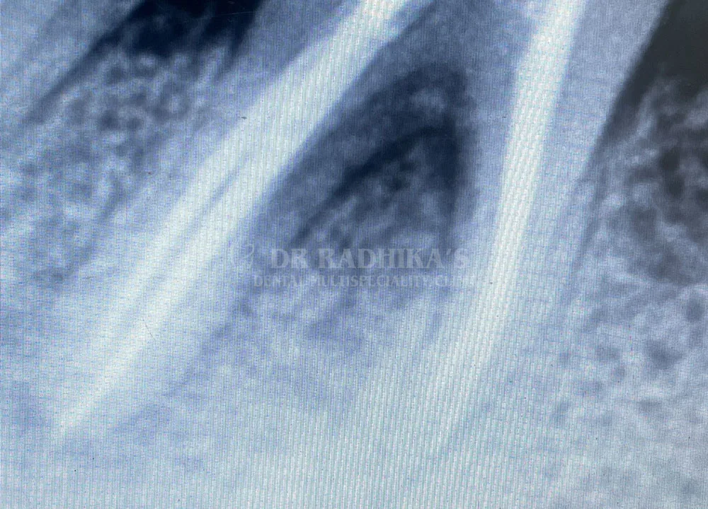
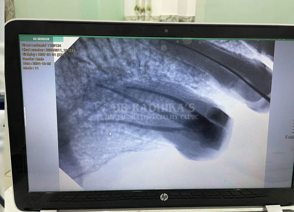
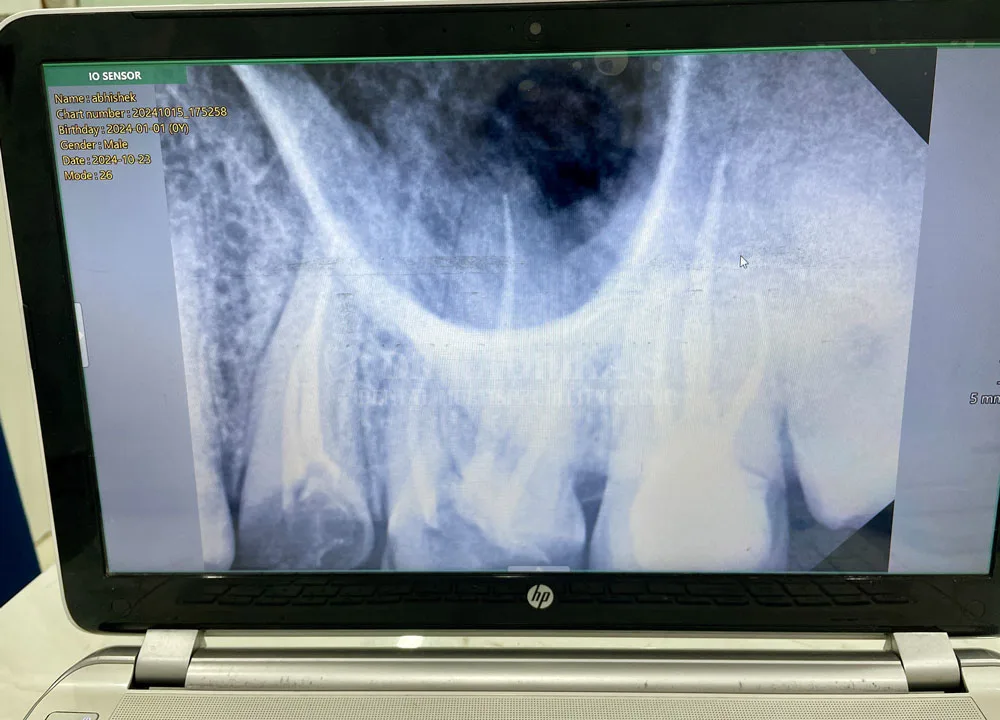
We also use digital X-rays and apex locators to ensure exact measurements and reduce radiation exposure.
The Procedure – Step-by-Step
Diagnosis and Imaging A thorough examination and digital X-ray are performed to assess the extent of the infection.
Access Opening A small opening is made in the crown of the tooth to access the pulp chamber.
Cleaning and Shaping The infected pulp is removed, and the root canals are cleaned, shaped, and disinfected under the microscope.
Filling and Sealing The cleaned canals are filled with a rubber-like material called gutta-percha and sealed to prevent future infection.
Restoration A crown or permanent filling is placed to restore the tooth’s structure and strength.
Your Best Smile Starts With Quality Expertise
Where Dentistry Meets Artistry. We create the most beautiful smiles and we keep those smiles beautiful.